
Summary
Persistent tooth pain after a root canal or vague, throbbing discomfort often points to hidden anatomy, such as micro-fractures or a missed accessory nerve canal. At Kingston Smiles NY, Dr. Ibrahim Sayeed, DDS, uses advanced, on-site 3D CBCT imaging to rotate and inspect your tooth structure in high definition. This eliminates the guesswork of traditional 2D X-rays, allowing us to pinpoint the precise source of infection and save your natural enamel safely.
Few things interfere with your daily life quite like chronic, unyielding tooth pain. You might feel a sharp sting when sipping hot coffee or a deep, lingering throb that keeps you awake at night. What makes this situation incredibly frustrating is when you visit a dentist, get a standard X-ray, and are told, “Everything looks completely normal.”
I’m Dr. Ibrahim Sayeed, DDS. Over my career, I have performed over 4,000 successful root canals, and I can tell you honestly: if a tooth continues to hurt, there is always an underlying physical reason. The issue isn’t your imagination; it is the limitation of traditional 2D imaging.
At Kingston Smiles NY, we specialize in solving mystery toothaches. By introducing state-of-the-art 3D CBCT imaging to our Kingston office, we can look deep inside your root anatomy to solve complex endodontic issues with absolute precision.
The Hidden Anatomy: What Standard 2D X-Rays Miss
Traditional dental X-rays are fundamentally two-dimensional. They display your teeth from a single flat angle, essentially functioning like a silhouette shadow on a wall. While excellent for detecting common surface cavities between teeth, a flat image is physically incapable of displaying what is hiding directly behind or deep within a dense root structure.
Human tooth root networks are incredibly intricate. They are rarely straight, simple, uniform tubes; instead, they frequently curve, split, or harbor tiny, microscopic offshoots known as accessory canals.
The “Missed Canal” Problem: If an auxiliary tooth root branch goes entirely untreated during a standard root canal procedure, active bacteria will remain trapped inside. This leads to a persistent, chronic infection that eats away at the jawbone structure surrounding the base of the tooth.
The 3D Precision Advantage
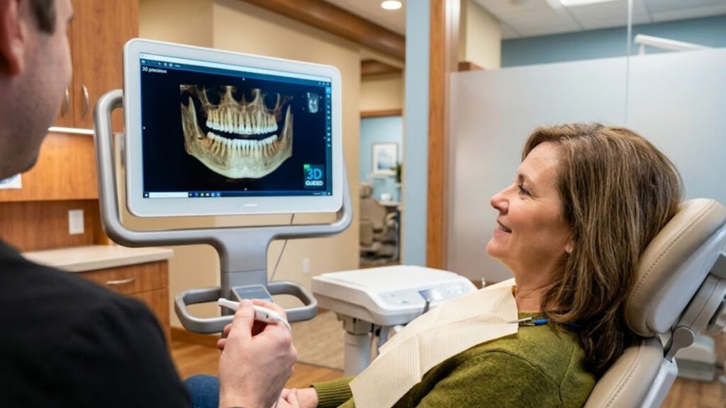
Our on-site CBCT (Cone Beam Computed Tomography) scanning equipment captures hundreds of individual structural data points in a single, comfortable 20-second open-air scan. This creates a pristine, highly detailed 3D digital model of your mouth. Using this model, I can digitally slice, cross-section, rotate, and zoom into your tooth’s interior pathways to map out every single microscopic canal before your treatment process ever begins.
3 Complex Endodontic Issues Solved by 3D Technology
Utilizing on-site 3D digital precision mapping directly transforms how we approach and execute complex root canals for patients traveling to see us from across Kingston, Saugerties, and Rhinebeck.
1. Identifying Microscopic Root Fractures
A vertical root fracture is a tiny crack that travels down the inside of a tooth root. These fractures are notoriously invisible on flat 2D films. With 3D imaging, we can rotate the tooth root on screen to detect these hidden cracks early, preventing you from undergoing unnecessary procedures if a tooth is structurally compromised.
2. Treating Calcified Canals
As we age, or in response to chronic trauma, dental canals can become “calcified”, meaning they narrow significantly and fill with hard mineral deposits. Attempting to navigate a calcified canal with traditional tools can lead to errors. A 3D scan acts as an absolute surgical GPS, showing me the exact path of the open canal down to the micrometer.
3. Resolving Previous Root Canal Failures (Retreatment)
If you had a root canal years ago at another dentist and it has started hurting again, it is likely due to a persistent infection at the very tip of the root. Our 3D bone mapping reveals the exact size and location of the infection, allowing us to perform a highly targeted retreatment to preserve your natural enamel.
Gentle, Technology-Driven Care in Kingston, NY
At Kingston Smiles NY, we combine this advanced dental technology with an empathetic, non-judgmental atmosphere. We know that the words “root canal” can cause immediate anxiety. By sitting down with you, displaying your 3D scan on our monitors, and explaining the step-by-step plan, we aim to replace your fear of the unknown with confidence.
Having this advanced diagnostic tool on-site at our 1105 Locust Street office means you do not have to travel to a separate imaging center or a distant specialist. We can diagnose and map out your path to relief right here during your consultation.
Reclaim Your Peace of Mind Today
You do not have to live with a chronic, mysterious toothache or a throbbing jaw. Experience the definitive diagnostic clarity and conservative, expert care designed to keep your natural smile fully intact.
Ready to find the definitive source of your tooth pain? Call our office today at 845-336-5855 or connect with our team online to reserve your comprehensive 3D Dental Assessment online today.
Practice Location
Our centralized office facility info is detailed below for seamless dental record routing:
- Address: 1105 Locust Street, Kingston, NY 12401 (Get directions on maps)
- Phone: +1 845-336-5855
- Email: kingstonsmilesny@gmail.com
- Office Hours: Tuesday and Wednesday: 8 AM – 5 PM
FAQ: Complex Root Canals & 3D Scanning
Why does my tooth still hurt months after a root canal?
This is typically caused by a hidden accessory canal that was missed during the initial treatment, an undetected micro-fracture in the root, or a new bacterial leak under your crown. A 3D CBCT scan is the most effective way to identify the exact cause.
Is a 3D dental scan safe?
Yes. Our modern CBCT machine focuses a highly targeted beam only on the necessary areas, utilizing up to 90% less radiation than a traditional hospital CT scan.
Does insurance cover 3D scanning for root canals?
Many PPO insurance plans (including local benefits like the Kingston Trust Fund and CSEA, as well as patient financing options like Cherry and CareCredit) cover 3D imaging when it is medically necessary to diagnose complex, persistent pain. Our administrative team will happily verify your benefits before your exam.